![]()
Medical Design Briefs - June 2023
Fighting Cancer with Real-Time Diagnostics
A Real-Time diagnostics solution has recently been cleared by the FDA. The results would allow clinicians intraprocedural identification and interpretation to properly assess tissue samples for adequacy. Developed by Aquyre Biosciences after a decade of research, CelTivity™ provides real-time cancer diagnostics in the operating room and private practice labs.
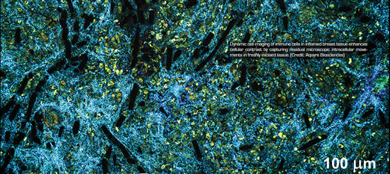
Dynamic cell imaging of immune cells inflamed breast tissue enhances cellular contrast by capturing residual microscopic intracellular movements in freshly excised tissue.
Inadequate real-time tissue assessment of biopsies from different cell types, like cancer cells, immune cells, granuloma, and others, forces proceduralists, such as bronchoscopists and radiologists, to choose between intraprocedural partial tissue adequacy assessment, rapid on-site evaluation (ROSE), or sending tissue samples for full pathology review. Neither truly answers the question, “Do we have enough cells to submit to pathology for the best chance of a conclusive diagnosis?” This can lead to prolonged delay for patient results, the need for a redo procedure, and potential delays for treatment options for the patient.
Consider lung biopsies, which are excised from patients and then sent to the laboratory for advanced cancer testing. These tests require that patients have an adequate amount of high-quality tissue to conduct advanced tests such as EGFR, kRas, or PD-1/PDL-1. To collect these biopsies, transthoracic needle biopsy (TTNA) is commonly used, which requires a needle to be inserted through the chest wall from outside the body. The risk of a collapsed lung (pneumothorax) can result in an average of 20 percent of procedures. Navigational bronchoscopy has emerged as a technology to collect lung biopsies with a lower risk of complications.
A better solution would be to assess tissue adequacy intra-procedurally, to interrogate and image the biopsy in its entirety, to determine: Do we have the tissue? What exactly are we looking at (cancer cells, immune cells, granuloma)? And, do we have enough cells to submit to pathology to gain the best chance of a conclusive diagnosis? The results would allow clinicians intraprocedural identification and interpretation to properly assess tissue samples for adequacy.
 Real-Time Diagnostics
Such a solution has recently been cleared by the FDA. Developed by Aquyre Biosciences, Inc. after a decade of research, CelTivity™ provides real-time cancer diagnostics of freshly excised cells and tissue in the operating room and private practice labs, effectively expanding advanced diagnosis capabilities.
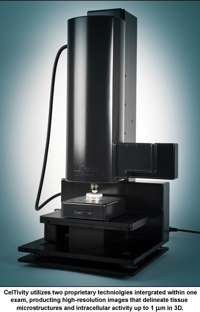
“Integrating digital images, data lakes, machine learning, and artificial intelligence, CelTivity utilizes two proprietary technologies integrated within one exam, producing high-resolution images that delineate tissue microstructures and intracellular activity up to 1 µm in 3D,” says Bertrand Le Conte de Poly, CEO of Aquyre Biosciences. “These technologies are full-field optical coherence tomography (FFOCT), and dynamic cell imaging (DCI). Of significance, is that these diagnostic results are available within two minutes. The freshly excised sample can then be sent to pathology, along with the CelTivity data. Moreover, tissue depth exploration can be achieved at 150 µm below the tissue surface with no cutting, staining, chemicals, or sectioning required.
Full-Field Optical Coherence Tomography
The CelTivity Full-Field OCT performs noninvasive, high-resolution optical slicing beneath the tissue surface. The full-field approach offers great potential in imaging of samples to assist fast diagnostic of cellular and morphological scale pathologies, such as cancer. It consists of an interference microscope, or more precisely, a classic Michelson interferometer with a microscope objective in each arm. The Michelson interferometer produces interference fringes by splitting a beam of light so that one beam strikes a fixed mirror and the other a movable mirror. When the reflected beams are brought back together, an interference pattern results.
An incoherent light source illuminates the whole field of the microscope objectives. Due to the low temporal coherence of the source, interference occurs only when the optical path lengths of the two arms of the interferometer are identical within 1 µm. When a biological sample is placed under the microscope objective in the sample arm, the light reflected by the reference mirror interferes with the light reflected or backscattered by the sample structures contained in a limited volume.
“The interferometer can be displaced to step the focal plane through different depths beneath the surface to create high-quality 3D tomographic images,” adds Le Conte de Poly. “Light-CT captures, en face, images directly using a megapixel camera and a pair of microscope objectives with lateral resolution to typically ~1 µm. The FFOCT image displays in gray scale the static optical contrast of the region of interest. Highly backscattering content like different fibrous structures (collagen, axons, etc.) appears white, while weakly backscattering content like cells appears dark, gray, or black.”
 Dynamic Cell Imaging
The CelTivity DCI enhances the cellular contrast by capturing the residual microscopic intracellular movements in freshly excised tissue. Dynamic cell imaging provides a dynamic contrast at the intracellular scale which complements the morphological information provided by FFOCT, thus bringing images up to the histology level of detail and information richness.
A DCI technique consists of recording the time series of each voxel - a unit of graphic information that defines a point in three-dimensional space - under examination during a few seconds, and processes them in order to extract characteristic features of the subcellular activity to be displayed. From a Fourier analysis of the time signal, the DCI RGB image is created where the blue channel corresponds to low frequency moving parts (< 0.5 Hz), the green channel displays medium frequency moving parts (0.5-5 Hz), and the red channel displays higher frequency moving parts (5-25 Hz). The signal intensity in each color channel reflects the dynamics amplitude in each corresponding frequency range.
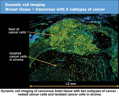
“DCI images obtained on different breast samples show the potential of the DCI technique to differentiate cancerous from normal tissue and ultimately to discriminate various cell types,” continues Le Conte de Poly.
Vibration Isolation
“Because CelTivity’s technologies function at the micron level, vibration isolation is critical to maintaining the system’s highest level of operation,” explaiins Le Conte de Poly. “Footfall from someone walking by, the closing of doors, HVAC units operating, or elevators in motion create low-frequency vibrations that can influence FFOCT and DCI imaging.”
“Consequently, Celtivity is supported by vibration isolation designed to more thoroughly cancel out low-frequency vibrations,” says Le Conte de Poly. “For this purpose, negative-stiffness vibration isolation was selected because of its high-performance capability, and ease of adaptability to hospital and laboratory environments.”
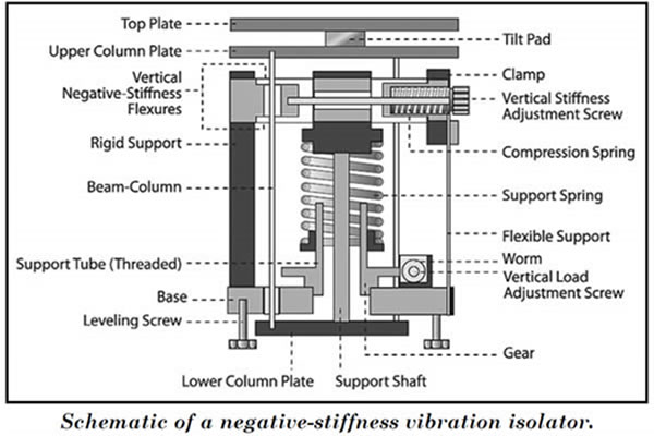
Negative-stiffness vibration isolation was developed by Minus K Technology, an OEM supplier to manufacturers of scanning probe microscopes, micro-hardness testers, and other vibration-sensitive instruments and equipment. These vibration isolators are compact and do not require electricity or compressed air, which enables sensitive instruments to be located wherever they are needed. There are no motors, pumps, or chambers, and no maintenance because there is nothing to wear out. They operate purely in a passive mechanical mode.
What is very advantageous about negative-stiffness isolators is that they achieve a high level of isolation in multiple directions. These isolators have the flexibility of custom tailoring resonant frequencies to 0.5 Hz vertically and horizontally (with some versions at 1.5 Hz horizontally). (Note that for an isolation system with a 0.5 Hz natural frequency, isolation begins at 0.7 Hz and improves with increase in the vibration frequency. The natural frequency is more commonly used to describe the system performance.)
Vertical-motion isolation is provided by a stiff spring that supports a weight load, combined with a negative-stiffness mechanism. The net vertical stiffness is made very low without affecting the static load-supporting capability of the spring. Beam-columns connected in series with the vertical-motion isolator provide horizontal-motion isolation. A beam-column behaves as a spring combined with a negative-stiffness mechanism. The result is a compact passive isolator capable of very low vertical and horizontal natural frequencies and high internal structural frequencies.”
Negative-stiffness isolators deliver very high performance, as measured by a transmissibility curve. Vibration transmissibility is a measure of the vibrations that are transmitted through the isolator relative to the input vibrations. Negative-stiffness isolators, when adjusted to 0.5 Hz, achieve approximately 93 percent isolation efficiency at 2 Hz; 99 percent at 5 Hz; and 99.7 percent at 10 Hz.
Better Image Quality
Celtivity’s technology is the sole technique providing axial resolution of 1 µm, which gives the highest quality image clarity and precision. It enables image capture on a 1 cm² region of tissue in one minute, thereby enabling immediate quality control of biopsies.
Full-field optical coherence tomography and dynamic cell imaging produce real-time images showing tissue architecture and intracellular activity data without sample processing or staining, and no need for dyes, contrast agents or tissue freezing.
This article was written by Jim McMahon, who writes on industrial, manufacturing, and technology issues. For more information on negative-stiffness isolators, contact Steve Varma, Minus K Technology, Inc.; Phone 310-348-9656; Fax 310-348-9638; e-mail request@minusk.com; www.minusk.com. For more information about Aquyre Biosciences, contact Bertrand Le Conte de Poly, CEO; Phone +33 1 78 96 50 00; e-mail info@ aquyre.com; www.aquyre.com.
|