![]()
May 2026
Minus K Educational Vibration Isolator Giveaway
2022 Winner Research Project
Optical Coherence Elastography - Pushing the Boundaries of Real-Time Strain and Elasticity Imaging of Biological Tissue
Optical coherence elastography (OCE) holds great promise for detecting and monitoring the altered mechanical properties of strain and elasticity with biological tissue that accompanies many clinical conditions and pathologies, particularly in cancer, cardiovascular disease and eye disease. Researchers at UC Irvine's Beckman Laser Institute are pushing the research envelope with OCE application, with the assistance of Negative-Stiffness vibration isolation.
by Jim McMahon
Tissue exhibits varying degrees of viscoelasticity (time-dependent response to a load), poroelasticity (presence of fluid-filled pores or channels), and anisotropy (a physical property that has a different value when measured in different directions), as well as a nonlinear relationship between elasticity and the applied load.
In establishing the link between elasticity and displacement, simplifying assumptions are usually made about tissue behavior and structure. Most commonly, that tissue is approximated as a linear elastic solid having mechanical properties which have the same value when measured in different directions (isotropic). Optical coherence elastography (OCE), however, has permitted a broader understanding regarding strain and elasticity in biological tissue.
OCE is a non-invasive imaging method for biological tissue, characterized by its niche in intermediate spatial resolution of tens to hundreds of micrometers, about one millimeter of depth penetration, and its high sensitivity to small mechanical changes at the microstrain level.
Optical Coherence Elastography
Elastography is a medical imaging technique used to measure tissue deformation under mechanical loads, enabling the mapping of local mechanical properties. The resulting images are known as elastograms. The term "elastography" has been in use since 1979, and significant advancements have been made in the field, primarily through ultrasound imaging, magnetic resonance imaging, and optical elasticity imaging.
Optical coherence elasticity imaging, one of the earliest optical elasticity methods, utilizes optical coherence tomography (OCT) to detect depth-resolved deformations in samples subjected to compression.
Displacement measurement plays a crucial role in OCE techniques, as tissue deformation often reveals essential mechanical properties. Although traditional OCT provides important diagnostic information, it is often inadequate for early diagnosis when structural deformations are minor.
Phase-sensitive detection is a primary method for detecting tissue deformation, similar to phase-based displacement detection in ultrasound imaging. This involves processing the phase-sensitive OCT signal to determine tissue displacement, followed by strain measurement. Compressional OCE integrates strain data from phase shift with stress applied through compression loading to calculate the elastic modulus, which characterizes the tissue's mechanical properties.
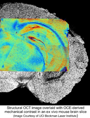
OCE presents new possibilities for various biomedical applications due to its superior resolution and mechanical sensitivity compared to ultrasound and magnetic resonance elastography. One particularly promising application lies in the differentiation between malignant and normal tissues, wherein OCE demonstrates superior contrast compared to conventional structural OCT imaging. There is a strong interest in accelerating OCE visualization for intraoperative use. By leveraging differences in the Young's modulus* of tumor components, OCE can produce images that closely resemble histological images. Unlike traditional histological techniques, which are invasive, time-consuming, and labor-intensive, OCE can be conducted on freshly resected tissue samples and even performed in vivo.
(*Young's modulus is a measure of the ability of a material to withstand changes in length when under lengthwise tension or compression).
OCE Research at UC Irvine's Beckman Laser Institute
 Researchers at UC Irvine's Beckman Laser Institute have considerable experience with OCE/OCT possibilities.
According to Fengyi Zhang, Ph.D Graduate Student with the Beckman Laser Institute, "We have focused on two destinations of OCE: a) Visualization of local movement and strains in biological tissue; and b) Visualization of elasticity of the tissue with mechanically produced deformations"
"The OCE technique is operable for a broad class of sufficiently soft biological tissues, even such tissues as cartilage," continued Dr. Zhang. "Previously, obtained strain and elasticity maps for such materials were obtained using mechanical testing. In contrast, OCE enables imaging in real-time."
In ophthalmology, OCE could be utilized to characterize the mechanical properties of the cornea to diagnose related ocular disease. With Dermatology, the elasticity of skin could indicate related pathologies. OCE technology could detect differences in stiffness of human skin layers in vivo. Oncological imaging ex vivo of excised tissues from different sample regions with different stiffness could be highlighted in a 2D depth-resolved elastogram. To improve management of atherosclerosis, OCE could be utilized to monitor the stability of plaque by mechanical characterization of the arterial wall.
Vibration Isolation
Maintaining the micron-level precision needed for the OCE technique at the Beckman
Laser Institute was compromised due to low-frequency vibrations originating from the building's air conditioning system, elevator movement and other facility operations.
"We were using an air table to help reduce these low-frequency vibrations, but with little success," explained Dr. Zhang. "Our data sets were being compromised."
"The problem was resolved however, when our laboratory was awarded a complementary Negative-Stiffness vibration isolation platform," said Dr. Zhang, in reference to being a winner of Minus K's Educational Vibration Isolator Giveaway .
Introduced in the mid-1990s by Minus K Technology, Negative-Stiffness vibration isolation has been widely accepted for vibration-critical applications, largely because of its ability to effectively isolate lower frequencies, both vertically and horizontally. The company's isolators are used by more than 300 universities and government laboratories in 53 countries.
Negative-Stiffness isolators are unique in that they operate purely in a passive mechanical mode. They do not require electricity or compressed air. There are no motors, pumps or chambers, and no maintenance because there is nothing to wear out.
"Vertical-motion isolation is provided by a stiff spring that supports a weight load, combined with a Negative-Stiffness mechanism," said Erik Runge, Vice President of Engineering at Minus K. "The net vertical stiffness is made very low without affecting the static load-supporting capability of the spring. Beam-columns connected in series with the vertical-motion isolator provide horizontal-motion isolation. A beam-column behaves as a spring combined with a negative-stiffness mechanism. The result is a compact passive isolator capable of very low vertical and horizontal natural frequencies and high internal structural frequencies."
Negative-Stiffness isolators achieve a high level of isolation in multiple directions, with the flexibility of custom-tailoring resonant frequencies to 0.5 Hz vertically and horizontally (with some versions at 1.5 Hz horizontally)*. When adjusted to 0.5 Hz, the isolators achieve approximately 93 percent isolation efficiency at 2 Hz, 99 percent at 5 Hz, and 99.7 percent at 10 Hz.
(*Note that for an isolation system with a 0.5 Hz natural frequency, isolation begins at 0.7 Hz and improves with increase in the vibration frequency. The natural frequency is more commonly used to describe the system performance.)
"The Negative-Stiffness isolation platform has considerably reduced the time and effort it takes us to process our OCE soft tissue research," explained Dr. Zhang. "The unit's portability and simplicity has been a substantial benefit to our lab."
Broad Possibilities
Added Dr. Zhang, "Overall, the achievements in the development of OCE-based methods of high-resolution elastography have already demonstrated rich unprecedented possibilities of this new modality for a broad range of biomedical applications.".
About the Beckman Laser Institute, Department of Biomedical Engineering, University of California, Irvine
The current research focus of the Beckman Laser Institute, Department of Biomedical Engineering, University of California, Irvine is on investigating light/tissue interactions; developing medical diagnostic and therapeutic devices and instruments using advanced optical, microfabrication and biomedical technologies; and applications of these technologies for the early diagnosis of disease.
For more information contact Fengyi Zhang, Ph.D Graduate Student with the Beckman Laser Institute, Department of Biomedical Engineering, University of California, Irvine, 1002 Health Sciences Road, Irvine California 92617; Phone 949-299-8277; email fengyiz1@uci.edu.
About Minus K Technology, Inc.
Minus K® Technology, Inc. was founded in 1993 to develop, manufacture and market
state-of-the-art vibration isolation products based on the company's patented negative-stiffness technology. Minus K products are used in a broad spectrum of applications including microscopy, nanotechnology, biological sciences, semiconductors, materials research, zero-g simulation of spacecraft, and high-end audio. The company is an OEM supplier to leading manufacturers of scanning probe microscopes, micro-hardness testers and other vibration-sensitive instruments and equipment. Minus K customers include private companies and more than 300 leading universities and government laboratories in 53 countries.
For more information on Negative-Stiffness isolators please contact Steve Varma,
Minus K Technology, Inc., 460 Hindry Avenue, Unit C, Inglewood, CA 90301 ? 310-348-9656 E-mail: sales@minusk.com Web: www.minusk.com
|